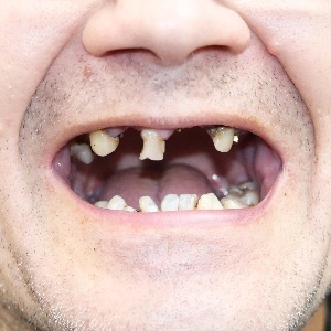
Being told you do not have enough bone for dental implants almost always means something narrower than it sounds. It means that one specific site in your jaw currently lacks the height, width, or density needed to anchor an implant safely — not that implants are permanently off the table for you. In most cases that deficiency can be measured precisely with a CBCT 3D scan and then either rebuilt with grafting or worked around using implant lengths, diameters, and angles that make use of the bone you still have. Bone loss is a clinical challenge, not a closed door.
One of the most common questions patients ask during a consultation is some version of: “Another office told me I’m not a candidate. Is that the end of it?” Usually it is not. What follows is a plain explanation of what that assessment actually measured, what it may not have measured, and what your real options are.
What Does “Not Enough Bone” Actually Mean?
An implant is an artificial tooth root. Like a natural root, it needs a surrounding envelope of living bone in every direction — above it, beside it, and around its full length. When a surgeon says there is not enough bone, they are describing a shortfall in one or more of three separate measurements. Those measurements fail independently, and confusing them is where a lot of patient anxiety comes from.
Height, Width, and Density Are Three Different Problems
- Height — The vertical distance between the top of the ridge and whatever structure sits beneath or above it. In the upper back jaw, the ceiling is the floor of the maxillary sinus. In the lower back jaw, the floor is the inferior alveolar nerve canal. Short height is an anatomical boundary problem, and it is the single most common reason patients in the upper molar region are turned away.
- Width — The thickness of the ridge from cheek side to tongue side. After a tooth is removed, the ridge narrows before it shortens, and the thin outer plate of bone resorbs first. A ridge can look perfectly adequate in height on a flat X-ray while being too thin to hold an implant without exposing threads.
- Density — How mineralized and structurally dense the remaining bone is. Density does not affect whether an implant fits; it affects whether the implant is stable the day it is placed and how it heals. The posterior upper jaw is naturally the softest bone in the mouth, which is why the same volume of bone behaves differently there than in the front of the lower jaw.
A patient can have generous height and unusable width. Another can have adequate volume in both directions and bone soft enough that a standard placement protocol would not be appropriate. These are different problems with different solutions, and they carry very different timelines.
Where the Bone Is Missing Changes the Plan
Location matters as much as quantity. Deficiency in the upper back jaw usually involves the sinus and is managed by lifting the sinus membrane and grafting beneath it. Deficiency in the lower back jaw is constrained by the nerve canal, which cannot be moved casually and often steers the plan toward shorter implants or vertical augmentation. Deficiency in the upper front is as much an appearance problem as a structural one, because the thin bone and gum over an implant in the smile zone determine whether the final crown looks natural.
If you want the background on why the bone disappeared in the first place — extraction, periodontal disease, long-term denture wear, prior implant failure — our overview of bone loss and dental implants covers the underlying biology in detail.
Why a Two-Dimensional X-Ray Cannot Answer This Question
A panoramic or periapical X-ray is a flat shadow of a three-dimensional structure. It gives a rough sense of height. It cannot show width at all, because width runs directly along the axis the image is projected through. It cannot reliably assess density. It carries magnification distortion that varies across the image, so a measurement taken from it is an estimate, not a number a surgeon can plan around.
Cone beam computed tomography solves this. A CBCT scan produces cross-sectional slices through the jaw, so bone can be measured in millimeters at the exact point where an implant would go. It maps the course of the nerve canal and the contour of the sinus floor. It allows density to be assessed at each proposed site. And the same dataset is used to design a surgical guide, so the plan made on screen is the plan carried out in the chair.
This is the honest crux of many second opinions: if the assessment you received did not include 3D imaging, it did not measure two of the three dimensions that decide the case. That is not a criticism of the clinician who saw you — it is a limitation of the tool that was available in that moment.
What Can Be Rebuilt, and Roughly How Long It Takes
Bone is one of the few tissues in the body that genuinely regenerates. Grafting does not glue a block of foreign material into your jaw permanently; it places a scaffold that your own bone grows into and gradually replaces. The category of procedures used to do this is advanced bone grafting, and the right one depends entirely on which dimension is short and where.
- Socket preservation — A graft placed into the socket at the time of extraction to limit the collapse that would otherwise occur. The least invasive option, and only available before the bone is lost.
- Ridge augmentation — Rebuilding width, height, or both along the crest of the jaw, usually with particulate graft material held in place by a barrier membrane, or with a solid block graft for larger defects.
- Sinus lift — Elevating the sinus membrane in the upper back jaw and placing graft material underneath it to create the height an implant needs.
- Growth factor adjuncts — Platelet-rich fibrin or plasma prepared from your own blood, used to support healing at the graft site. These improve the environment; they do not replace graft volume.
Timelines vary by technique, defect size, and how you heal. Straightforward grafts commonly integrate over a period of a few months before an implant can be placed; larger reconstructions in the literature are commonly cited in the range of six to nine months. Some smaller defects allow the graft and the implant to be done in a single surgery. A surgeon should be able to tell you which category your case falls into after reviewing a scan, and should tell you the range honestly rather than the best-case number.
Options That Reduce or Skip Grafting Altogether
Grafting is not the only path, and for some patients it is not the best one. Several approaches make use of existing bone instead of building new bone.
- Short implants — Modern implant surfaces allow shorter fixtures to perform predictably in situations that once required added height. Where height is limited but width and density are good, a shorter implant may avoid a sinus lift entirely.
- Tilted or angled placement — Angling implants to engage the denser bone that remains at the front of the jaw, avoiding the deficient posterior regions. This is the principle behind full-arch protocols that support a fixed bridge on four or six implants.
- Narrow-diameter implants — Useful in thin ridges and tight spaces, with the tradeoff that they distribute force differently and are not appropriate for every position.
- Zygomatic implants — For severe upper-jaw atrophy, long implants anchored in the cheekbone rather than the maxilla. A demanding procedure reserved for specific cases, but one that can bypass years of staged grafting.
Each of these trades something. Avoiding a graft usually means accepting a narrower margin for error in placement, which is precisely why these approaches depend on planning quality and surgical experience rather than on the technique alone.
When a “No” From One Practice Is Not the Final Answer
Most general dentists place a modest number of implants each year, and a case involving significant bone deficiency sits well outside routine work. Declining it is a reasonable and responsible decision. It is not the same thing as the case being untreatable — it means the case belongs in a setting where reconstruction is everyday work. We write about this distinction in more detail in why not every implant case is simple.
A genuine re-evaluation should include all of the following:
- A new or reviewed CBCT scan with site-specific measurements you can see on screen
- Identification of the cause of the bone loss and whether it is still active
- A review of medical history and medications that affect bone healing
- At least two alternative plans — typically one that grafts and one that avoids grafting — with the tradeoffs of each stated plainly
- A realistic timeline and sequence, including how you will eat and look during the interim
What to Bring to a Second-Opinion Consultation
You will get a far more useful hour if you arrive with the following:
- Any imaging already taken — including CBCT data on a disc or file, which avoids repeat radiation and lets a new surgeon measure the same anatomy
- A written list of medications and supplements, particularly bone-density drugs, blood thinners, steroids, and anything taken for autoimmune conditions
- Your medical history, including diabetes control, any history of radiation to the head or neck, and smoking status
- What you were actually told, in as much detail as you remember, and any treatment plan or estimate you were given
- Your own goals — a fixed bridge, a single tooth, an end to a loose denture — and your real constraints on time and travel
The reason this level of preparation pays off is that reconstructive implant planning is a judgment discipline, not a checklist. Our discussion of why a specialty implant practice matters explains how case volume and in-house imaging change what is possible to plan for.
When “No” Really Is No
Honesty in both directions builds trust, so here is the other side. There are situations where implants are genuinely inadvisable, at least for now:
- Active, untreated infection — Advanced periodontal disease or a persistent site infection must be resolved and stabilized before any graft or implant is placed. This is a sequencing problem more often than a permanent one.
- Certain medications — Intravenous antiresorptive therapy and some other bone-modifying drugs meaningfully change surgical risk. This requires coordination with your physician, and the decision belongs with them, not with a website.
- Uncontrolled systemic conditions — Poorly controlled diabetes and some autoimmune or healing disorders can make graft failure likely enough that surgery is not the right recommendation until the condition is managed.
- A history of high-dose radiation to the jaws — This alters bone healing capacity in ways that require specialized evaluation and sometimes rules out grafting in the irradiated field.
- Continued heavy smoking — Smoking constricts the blood supply that a graft depends on. This is the one item on the list a patient can change.
Even in these cases, the honest answer is usually “not this, not now” rather than “nothing ever.” A well-made implant-retained overdenture, a conventional prosthesis, or a staged plan that begins with medical management may be the better recommendation. This article is general education, not medical advice, and any decision involving your medications or systemic health should be made together with your physician and your surgeon.
Frequently Asked Questions
How much bone do you actually need for a dental implant?
There is no single number, because the requirement depends on the implant chosen and the site. In general terms, a surgeon looks for enough width to leave sound bone on both sides of the implant and enough height to stay clear of the sinus or nerve. What matters more than the raw figure is whether the available bone can be engaged safely at the angle the final restoration requires.
Can bone grow back on its own after tooth loss?
No. Once alveolar bone resorbs after a tooth is lost, it does not regenerate spontaneously, because the stimulation that maintained it is gone. It can be rebuilt surgically with grafting, and the earlier that is done, the smaller the reconstruction tends to be. This is the reasoning behind placing a graft at the time of extraction rather than waiting.
Is bone grafting painful?
Most patients describe graft recovery as comparable to an extraction — soreness and swelling that peak in the first two or three days and then steadily improve, managed with prescribed medication and cold compresses. Larger reconstructions involve more swelling and a longer soft-tissue recovery. Pain that worsens after the first week rather than improving is the pattern worth reporting promptly.
Do I have to wait months before I have teeth again?
Waiting for a graft to integrate does not mean going without teeth. Interim solutions — a temporary partial, an existing denture adjusted to avoid loading the graft site, or a provisional bridge — are planned as part of the treatment sequence. In some cases an implant can be placed at the same visit as the graft, shortening the overall timeline considerably.
Get a Straight Answer About Your Own Bone
If you have been told implants are not possible, the most useful next step is a scan and a conversation with a surgeon who reconstructs deficient jaws routinely. You may hear the same answer. You may hear a different one. Either way you will know what your anatomy actually looks like and why. You can schedule a complimentary implant consultation to have your situation evaluated in three dimensions.
Center for Dental Implants welcomes patients from across South Florida, with offices in Aventura, Hallandale Beach, and Pembroke Pines.