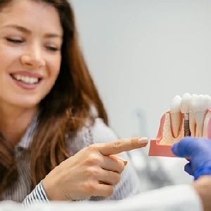
Peri-implantitis is inflammation around a dental implant that has progressed past the gum tissue and into the bone supporting the implant, producing measurable bone loss over time. It is not the same as peri-implant mucositis — inflammation confined to the soft tissue, which is generally reversible when it is caught and treated early. And it is not the same as a failed implant. An implant with peri-implantitis is usually still integrated in bone and still functioning; the disease threatens that integration rather than having already ended it. Treatment ranges from non-surgical cleaning and decontamination to surgical access, implant surface treatment and regenerative grafting — though in advanced cases, removing the implant may still be the right decision.
One of the more unsettling conversations patients have with us begins with some version of: “They told me I have peri-implantitis. Does that mean I’m losing the implant?” Often, the honest answer is no — not yet, and not necessarily. But the answer depends entirely on how much bone has already been lost, how fast it is being lost, and what is driving the process. That assessment requires an in-person examination and current imaging. It cannot be made from a description of symptoms.
What Is Peri-Implantitis, Exactly?
A dental implant is not a tooth. A natural tooth is suspended in bone by the periodontal ligament, a fibrous attachment with its own blood supply and a degree of immune surveillance. An implant has no ligament. It is fused directly to bone — osseointegrated — and sealed at the top only by a cuff of soft tissue that adheres to the implant or abutment surface.
That soft-tissue cuff is the entire biological barrier between the mouth and the bone holding the implant. It is a reasonable barrier, but it is less robust than the attachment around a natural tooth, and it has less capacity to wall off a bacterial front once one establishes itself.
Peri-implantitis is what happens when bacterial biofilm accumulates at that junction, the tissue becomes inflamed, and the inflammation extends apically — downward — into the bone. The body’s own inflammatory response resorbs bone in an attempt to move away from the bacterial insult. The result is a progressive, crater-shaped defect around the implant that shows up on radiographs as bone level dropping year over year.
Two points matter here. First, the process is driven by bacteria and by the host’s response to them — it is a biological disease, not simply mechanical wear. Second, it is progressive, which means the earlier it is interrupted, the more bone there is left to work with.
Peri-Implant Mucositis vs Peri-Implantitis: The Distinction That Changes Everything
These two terms get used interchangeably in conversation, and they should not be. They describe different stages of the same biological process, and they carry very different prognoses.
Peri-implant mucositis is inflammation of the soft tissue around an implant with no bone loss beyond the small amount of remodeling that normally occurs in the first year after placement. The gum is red, it may be puffy, and it bleeds when probed. The bone underneath is intact.
Peri-implantitis includes those soft-tissue signs but adds progressive bone loss around the implant body. That is the defining difference, and it is why an accurate diagnosis depends on radiographs, not on how the gum looks.
| Factor | Peri-Implant Mucositis | Peri-Implantitis |
|---|---|---|
| Tissue involved | Soft tissue only | Soft tissue plus supporting bone |
| Bone loss on radiographs | None beyond normal early remodeling | Progressive loss compared with earlier films |
| Bleeding on probing | Typically present | Typically present, sometimes with pus |
| Probing depths | Usually stable | Increasing over time |
| Implant mobility | None | None until very late; mobility is a grave sign |
| Reversibility | Generally reversible with prompt treatment and improved hygiene | Can often be arrested; lost bone does not return on its own |
| Typical first-line care | Professional debridement, hygiene coaching, closer recall | Debridement first, then surgical access if the defect persists |
The clinically useful way to hold this: mucositis is the window in which the problem is most straightforward to reverse. Peri-implantitis is the stage at which the goal shifts from reversal to arrest — stopping further loss and, where the defect anatomy allows, attempting to rebuild some of what was lost.
Why Peri-Implantitis Isn’t the Same as a Failed Implant
A failed implant is one that has lost osseointegration. It is mobile, it is no longer anchored, and it needs to come out. That is a different clinical situation entirely.
An implant with peri-implantitis is, in most cases, still solidly integrated along the portion of its length that remains in healthy bone. It does not move. It still supports a crown. What it has is an active disease process eating away at its foundation from the top down.
This distinction matters because it determines what the treatment is trying to accomplish. In a failed implant, the conversation is about removal, site healing and reconstruction. In peri-implantitis, the conversation is about stopping a disease while the implant is still salvageable. Those are different conversations, and conflating them can lead patients to give up on an implant that still has a reasonable chance — or, in the other direction, to assume they have more time than they do.
Being told you have peri-implantitis is a call to act, not a verdict. But it is a genuine call to act. Bone that is lost while a decision is being deferred is bone that has to be rebuilt later, if it can be.
What Raises the Risk of Peri-Implantitis?
Peri-implantitis rarely has a single cause. It usually reflects several factors stacking up around one implant site.
- Plaque accumulation and poor cleaning access — The most direct driver. Biofilm at the implant–tissue junction initiates and sustains the inflammation. If the patient cannot physically reach the area with a brush, floss or interdental aid, plaque control fails no matter how motivated they are.
- Prosthesis design that blocks cleaning — An over-contoured crown, a bridge with insufficient space beneath it, or a full-arch prosthesis with a tight tissue interface can make adequate hygiene mechanically impossible. This is a design issue, and it is one of the more correctable contributors.
- Residual cement — When a crown is cemented onto an abutment, excess cement can be pushed below the gum line and left behind. Cement acts as a persistent irritant and a surface for biofilm. It is a well-recognized trigger and one reason screw-retained restorations are often preferred where the case allows.
- A history of periodontitis — Patients who lost natural teeth to gum disease carry a bacterial profile and a host inflammatory response that predispose them to the same process around implants. Stabilizing periodontal health is central to protecting implants, and our periodontal and gum care protocols exist in large part for this reason.
- Smoking — Nicotine constricts blood vessels and impairs the soft-tissue seal and healing capacity around implants. Smoking is consistently identified in the literature as a significant risk factor.
- Uncontrolled diabetes — Poor glycemic control impairs wound healing and immune response. Well-controlled diabetes is a far smaller concern than uncontrolled diabetes. This is a question for your physician as much as your surgeon, and medical coordination matters.
- Lack of maintenance — Implants that are never professionally monitored can lose meaningful bone before anyone looks. Peri-implantitis is largely painless in its earlier stages.
This is general education rather than medical advice. Which of these factors is actually operating in your case is something that can only be determined by examining you and reviewing your records.
How Is Peri-Implantitis Diagnosed?
A single photograph or a single X-ray does not diagnose peri-implantitis. Diagnosis rests on a combination of findings, and crucially on change over time.
- Probing depths — A calibrated probe is walked around the implant to measure how deep the sulcus is. Increasing depth relative to a prior baseline is a meaningful signal.
- Bleeding and suppuration on probing — Bleeding indicates active inflammation. Pus indicates a more established infectious process.
- Radiographic bone level over time — This is the pivotal measurement. The clinician compares the current bone level against the earliest available film — ideally the one taken at the time the restoration was delivered. A single snapshot showing a certain bone level cannot tell you whether that level is stable or actively dropping.
- Three-dimensional imaging — CBCT imaging shows the defect in three dimensions, including whether bone is missing on the facial or lingual side, which a flat film cannot reveal. Defect shape strongly influences whether regeneration is realistic.
- Assessment of the restoration — The crown or bridge is examined for contour, fit, retained cement and access for cleaning.
This is why bringing your old radiographs to any evaluation is genuinely useful. Without a prior reference point, a clinician is left estimating.
The Non-Surgical Treatment Ladder
Treatment begins with the least invasive interventions that have a reasonable chance of controlling the disease.
- Mechanical debridement — Removal of biofilm and calculus from the exposed implant surface using instruments chosen not to scratch or contaminate titanium — titanium or ceramic-tipped scalers, air-polishing with fine powders, or ultrasonic tips designed for implants.
- Surface decontamination — Chemical or mechanical treatment of the exposed threads to disrupt the biofilm that mechanical instruments cannot fully reach. Implant surfaces are microscopically textured to encourage bone attachment, and that same texture shelters bacteria.
- Local antimicrobials — Antiseptic irrigation or locally delivered antimicrobial agents may be used alongside debridement in selected cases.
- Correcting the contributing factor — Removing retained cement, modifying or remaking an unhygienic prosthesis, or reshaping a crown that traps plaque. If the cause is not addressed, the disease returns.
- Hygiene instruction and shortened recall — Specific tools for the specific site, and more frequent professional monitoring.
Non-surgical therapy alone is more predictable in mucositis than in established peri-implantitis. In deeper defects, instruments simply cannot reach the full implant surface through a closed pocket. When re-evaluation shows persistent bleeding, persistent depth, or continued bone loss, the next step is surgical access.
When Surgery Becomes the Right Step
Surgical treatment is not a last resort. It is often the point at which the disease can finally be addressed directly, because the implant surface becomes visible.
- Access flap surgery — The gum is reflected to expose the defect and the affected implant threads, allowing thorough cleaning under direct vision.
- Implant surface decontamination — With the surface exposed, more complete mechanical and chemical decontamination becomes possible.
- Implantoplasty — Smoothing and polishing exposed threads so the surface no longer harbors biofilm. This is generally reserved for portions of the implant that will remain exposed rather than being regenerated over.
- Regenerative grafting — Where the defect is contained — a crater with intact bone walls around it — bone grafting material, often with a barrier membrane and biologic adjuncts, may be placed in an attempt to rebuild support. Contained defects respond more predictably than broad, unwalled defects.
- Resective approaches — In some anatomies, reshaping the bone and repositioning the tissue to create a cleanable architecture is more realistic than trying to regenerate.
Which approach fits depends on the shape of the defect, the position of the implant, the thickness and quality of the surrounding tissue, and the patient’s ability to maintain the result. Regeneration is attempted where the anatomy supports it — it is not universally achievable, and no responsible surgeon can promise a specific amount of bone fill.
When Removal Becomes the Right Call
Some implants should come out. Stating this plainly is more useful than optimism.
Removal generally becomes the appropriate recommendation when bone loss has advanced past the point where remaining support is meaningful, when the implant has become mobile, when the implant is fractured, when the implant was placed in a position that cannot be restored or cleaned, or when repeated treatment has failed to arrest the disease.
Explantation is not a failure of care — it is a clinical decision that resets the site. In many cases the site can be grafted and, after healing, a new implant placed in a better position with a better-designed restoration. That sequence is the substance of implant rescue and revision treatment, and it is frequently more technically demanding than the original placement was.
Why Bone Grafting Expertise Matters So Much Here
Regenerative peri-implantitis treatment is, at its core, a bone grafting problem performed in a compromised, previously infected, hard-to-access site. The defect is irregular. The surrounding tissue is often thin and scarred. The graft has to integrate against a titanium surface rather than against a clean bony wall. Soft-tissue closure over the graft has to be tension-free or the whole effort is undermined.
These are exactly the variables that separate a routine graft from a difficult one. Dr. Arun K. Garg spent over 20 years as a full-time professor of surgery in the Division of Oral and Maxillofacial Surgery at the University of Miami Leonard M. Miller School of Medicine, where he also served as director of residency training. He is considered the world’s preeminent authority on bone biology, bone harvesting and bone grafting for dental implant surgery, has published nine books translated into multiple languages, and has trained more than 20,000 dentists. When a case turns on whether a defect can be rebuilt, that depth of grafting judgment is the variable that matters most.
The Maintenance Protocol That Prevents Recurrence
Treatment stabilizes a site. Maintenance keeps it stable. Peri-implantitis recurs when the conditions that produced it return.
- Professional maintenance on a defined interval — Often more frequent than standard six-month recall for patients with a history of peri-implant disease. The interval should be set by risk, not by habit.
- Probing and radiographic monitoring at each interval — Documented, so that change over time is visible rather than inferred.
- Daily hygiene matched to the prosthesis — Interdental brushes, water flossers, superfloss or specific threaders, chosen for the actual geometry in your mouth.
- A prosthesis you can actually clean — If the restoration blocks access, no home-care routine will compensate. Redesigning it is sometimes the single most valuable intervention.
- Managing systemic and behavioral risk — Smoking cessation support and coordination with your physician on glycemic control, where relevant.
Frequently Asked Questions
Can peri-implantitis be cured?
It is more accurate to speak of arresting peri-implantitis than curing it. In many cases, treatment stops further bone loss and returns the tissue to a healthy, non-bleeding state that can be maintained. Bone already lost does not regrow on its own, though regenerative grafting can sometimes rebuild part of it. In advanced cases, the disease cannot be arrested and the implant needs to be removed.
Does peri-implantitis hurt?
Often it does not, particularly in the earlier stages, which is precisely why it can progress unnoticed. Some patients notice bleeding when brushing, a bad taste, tenderness, or gum that looks red or has receded around the crown. Discomfort or pus tends to appear later. Because pain is an unreliable indicator, radiographic monitoring at maintenance visits does the detection work.
How fast does peri-implantitis progress?
The rate varies considerably between patients and even between implants in the same mouth. Some sites lose bone slowly over years; others progress more rapidly, particularly where smoking, uncontrolled diabetes or a history of aggressive periodontitis is involved. Because the rate cannot be predicted from a single visit, comparing sequential radiographs is the only way to know how a specific site is behaving.
If I lose one implant to peri-implantitis, will I lose the others?
Not necessarily. Peri-implantitis is site-specific as well as patient-specific — a local factor such as retained cement or an uncleanable crown may explain one site entirely. That said, one affected implant is a reason to evaluate every implant you have and to reassess overall risk factors, since some contributors act throughout the mouth.
Get a Clear Picture of What Is Actually Happening Around Your Implant
Peri-implantitis is treatable more often than patients are led to expect, and the earlier it is addressed the more options remain. What it is not is something that can be assessed from a description — it requires an in-person examination, probing, and current imaging compared against your earlier films. If you have been told you have bone loss around an implant, or you have noticed bleeding or recession around a crown, a consultation and evaluation will tell you where you actually stand.
Center for Dental Implants welcomes patients from across South Florida, with offices in Aventura, Hallandale Beach, and Pembroke Pines.