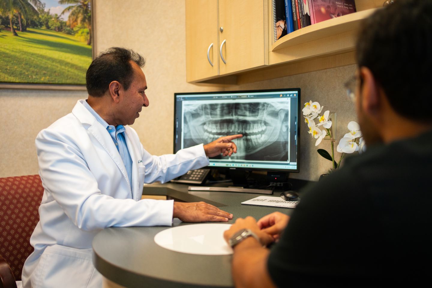
A CBCT scan is a three-dimensional X-ray of your jaw, and it shows the things a flat dental X-ray physically cannot: how wide your bone is at the exact spot an implant would go, how dense that bone is, precisely where the nerve canal runs, where the sinus floor sits, and what is left behind from old extractions or a failed implant. A standard panoramic or periapical film compresses all of that into a single flat image, so it can show bone height but never bone width or the true position of anatomy front-to-back. Implant surgery is a three-dimensional problem, and the scan is what turns guesswork about your specific anatomy into measurable planning.
What Is a CBCT Scan?
CBCT stands for cone beam computed tomography. The machine rotates a cone-shaped X-ray beam once around your head while a sensor captures hundreds of images from every angle. Software then reconstructs those images into a three-dimensional volume of your jaw that can be sliced and viewed from any direction — from the side, from above, and in cross-section through a single tooth site. It is not the same machine as a medical CT scanner. CBCT was developed specifically for the head and jaws, and it produces high-resolution images of hard tissue at a considerably lower radiation dose than a conventional medical CT of the same region.
Why Isn’t a Regular Dental X-ray Enough?
This is the part that surprises patients most, and it is worth being precise about. A panoramic X-ray or a periapical film is a two-dimensional projection of a three-dimensional structure. Everything in the beam’s path is flattened onto one plane.
The practical consequences are specific:
- You see height, not width — A flat film can suggest there is plenty of bone from the top of the ridge down to the nerve. It cannot tell you whether that ridge is a comfortable ten millimeters wide or a knife-edge three millimeters wide. Ridges frequently resorb in width first.
- Structures overlap — Roots, sinus walls, and the nerve canal all superimpose on one another. What looks like a clear space may be two structures stacked in the beam.
- Distances are distorted — Panoramic images magnify unevenly across the arch, so a measurement taken from the film is an approximation, not a true dimension.
- Position front-to-back is invisible — You cannot tell whether the nerve canal runs along the tongue side or the cheek side of the planned implant, which is precisely the information that determines a safe drilling angle.
None of this makes 2D imaging obsolete. It remains the right tool for checking a filling, monitoring a root, or screening the whole arch quickly. It is simply the wrong tool for planning surgery in bone you cannot see.
2D Dental X-ray vs 3D CBCT
| Question | 2D Panoramic or Periapical X-ray | 3D CBCT Scan |
|---|---|---|
| Bone height at the site | Approximate, with magnification error | Measured accurately in millimeters |
| Bone width at the site | Not visible at all | Visible in cross-section and measurable |
| Bone density and quality | Only a general impression | Assessable slice by slice |
| Nerve canal position | Rough vertical location only | Full course traced in three dimensions |
| Sinus floor and sinus health | Outline only; pathology easily missed | Floor contour, septa, and lining changes visible |
| Defects from prior surgery | Often obscured by overlap | Shape and extent of the defect defined |
| Can it drive a surgical guide? | No | Yes — it is the required starting data |
| Radiation dose | Very low | Higher than a single film, low relative to medical CT |
What Does the 3D Scan Actually Reveal?
- Bone width and height at the exact planned site — Not an average for the region, but the real dimensions of the specific millimeter of ridge where the implant would sit.
- Bone density and quality — Dense cortical bone and soft trabecular bone behave very differently under a drill and during healing. Knowing which you are working with changes drilling protocol, implant selection, and whether a temporary tooth can be attached the same day.
- The inferior alveolar nerve canal — In the lower jaw this canal carries the nerve supplying sensation to the lip and chin. Its vertical position, its side-to-side course, and how far it loops forward all vary between patients. The scan lets that path be traced and respected.
- The sinus floor and any sinus pathology — In the upper back jaw, the maxillary sinus limits available height. The scan shows the floor’s true contour, any bony walls dividing the sinus, and thickening of the lining that may need attention before grafting.
- Existing root positions and angulation — Neighboring roots sometimes lean into the space where an implant needs to go. That is far easier to see in cross-section than on a flat film.
- Defects from prior extractions or failed implants — Where bone is missing, the scan defines the shape of the void, which determines what kind of graft is appropriate.
Why This Matters Most in the Cases We See
For a straightforward single implant in generous bone, a scan confirms what was already likely. Its value rises sharply in the cases that make up much of our practice.
When a ridge has resorbed, the entire question is how much and in what direction — a deficiency in width calls for a different approach than a deficiency in height, and only a 3D view distinguishes them. That measurement is what shapes an advanced bone grafting plan rather than a general intention to “add some bone.” When the sinus is the limiting factor, the scan determines whether a modest crestal lift will do or whether a lateral approach is required, and it flags the anatomy — thin membranes, dividing septa, existing sinus inflammation — that makes sinus grafting more demanding. And in revision cases, where a previous implant did not integrate, the scan is the only way to see the true shape of the defect left behind before deciding how to rebuild it.
What Is the Scan Like for You?
Short and undramatic. You sit or stand at the machine with your chin on a small rest and a strap or bite piece to keep your head still. The arm rotates once around your head. The capture itself takes a matter of seconds. There is no tunnel, no enclosure, and nothing touching your face beyond the chin rest — patients who find medical CT or MRI confining are usually surprised by how open it is.
On radiation, the honest framing is comparative rather than numerical. A CBCT delivers more dose than a single dental X-ray and substantially less than a medical CT scan of the same area. It is ordered when the information genuinely changes the surgical plan, which for implant surgery it almost always does — and the alternative, operating with incomplete information about nerve and sinus position, carries its own risk. If you are pregnant or think you may be, tell us before the scan is taken.
How the Scan Becomes a Surgical Plan
The scan is data, not a decision. What happens next is where the value is realized:
- The volume is loaded into planning software where the nerve canal is traced and the sinus floor mapped.
- A digital scan of your teeth is merged with it, so bone anatomy and the shape of your bite exist in the same model.
- The final tooth is designed first — its position, angle, and how it will meet the opposing teeth.
- The implant is then positioned virtually to support that tooth, at a diameter, length, and angle the available bone can accept.
- A surgical guide is 3D printed from that plan — a physical template that seats over the teeth or ridge and directs the drill along the planned path.
Every one of those steps depends on the accuracy of the original volume, which is why the imaging and planning tools a practice uses are not an incidental detail. You can see the equipment behind this workflow on our technology page.
What a Flat X-ray Can Miss: A Common Pattern
Consider a familiar scenario, described generally rather than as any individual patient. A lower back tooth was removed several years ago. A panoramic film shows what looks like a healthy span of bone above the nerve canal — comfortably more than enough height for a standard implant. On that basis, the case reads as routine.
The CBCT tells a fuller story. In cross-section, the ridge has resorbed from the cheek side inward and is now narrow at the crest, tapering to a thin edge. There is height, but not enough width to house an implant with bone remaining on both sides. The scan may also show the nerve canal sitting toward the cheek side rather than centered, further narrowing the safe corridor.
Nothing here was hidden through anyone’s oversight — the information simply was not present in a two-dimensional image. What changes is the plan: instead of a single surgery, the case becomes ridge augmentation first, healing, then placement into rebuilt bone. Finding that out before surgery is a very different experience from finding it out during surgery.
Frequently Asked Questions
Do I need a CBCT for every dental implant?
Most implant cases benefit from one, and current practice strongly favors 3D imaging before surgical placement. The scan is ordered when it will meaningfully affect the plan — which is nearly always true when an implant is going into bone whose width, density, and relationship to the nerve or sinus cannot be confirmed any other way.
How long does a CBCT scan take?
The scan itself takes seconds, and the full appointment step usually runs a few minutes including positioning. Images are available immediately, so it is common to take the scan and review it with you on screen during the same visit rather than asking you to return.
Is a CBCT the same as a medical CT scan?
No. A medical CT uses a fan-shaped beam and an enclosed gantry, and delivers a considerably higher radiation dose. CBCT uses a cone-shaped beam in an open machine designed for the jaws, producing detailed hard-tissue images at a fraction of that dose.
Can I bring a CBCT scan taken somewhere else?
Often, yes. Bring the image files rather than printouts, since the surgical value lies in the full volume that can be sliced and measured. Whether it can be used depends on how recent it is, how much of the jaw it captured, and its resolution — an older or partial scan sometimes needs to be repeated.
See Your Own Anatomy Before Anyone Decides Anything
Reading your scan together is usually the moment the plan stops feeling abstract. You see where the bone is, where it is not, and what would have to happen to place an implant safely. If you have been given an answer about implants without 3D imaging behind it, that is a reasonable point to seek a second look. Schedule a consultation and we will walk through the images with you.
Center for Dental Implants welcomes patients from across South Florida, with offices in Aventura, Hallandale Beach, and Pembroke Pines.